We are experiencing a very high volume of calls and messages and ask for your patience. We will answer your portal messages within 48 hours.
We are experiencing a very high volume of calls and messages and ask for your patience. We will answer your portal messages within 48 hours.

Embryo biopsy is a normal part of preimplantation genetic diagnosis (PGD), which can be used to select "normal" embryos for transfer. Historically, biopsy was done on day-3 embryos (at the 8-cell stage). Day-3 biopsy is now rarely done for the following reasons: a) there was a relatively high chance of misdiagnosis due to "aneuploidy" (the one cell that was biopsied did not represent the remaining seven) and b) a decrease in pregnancy rates due to manipulation of the embryo.
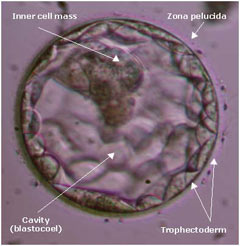
Currently, embryo biopsy for PGD is done on blastocysts (day-5 or 6 after fertilization). To understand how and why this is done, we should first get familiar with the anatomy of a blastocyst (see figure below).
 A blastocyst consists of three parts: a) The inner cell mass, which forms the baby, b) the outer cell mass or trophectoderm, which forms the placenta, and 3) a fluid filled cavity -- the blastocele. These are surrounded by the zona pellucida (the egg shell).
Trophectoderm biopsy has become the preferred method of obtaining cells for use in PGD. Since multiple cells are analyzed, the results are more accurate. Also, there is no decrease in pregnancy rates when a blastocyst is biopsied. Another advantage that patients feel strongly about is the fact that the biopsy is taken from the part of the embryo that forms the placenta (trophectoderm or outer cell mass). The inner cell mass (that forms the baby) remains untouched!
The process of obtaining those cells has been made easier by the use of laser technology. The laser is used to make a breech in the zona pellucida (ZP) of the embryo on day-3. The technique used is similar to what is done for assisted zona hatching (AZH; a technique, which can help with implantation).
As the embryo divides and cell numbers increase, the trophectoderm cells herniate out through the breech made in the ZP, and these cells can be biopsied without detriment to the embryo.
When the trophectoderm cells herniate out of the zona, a small sample of those cells are biopsied for use in determining the genetic constitution of the embryo.
The biopsy is performed by anchoring the embryo to what is called a ‘holding pipette”. This pipette uses gentle suction to hold the embryo in place while a second pipette, the biopsy pipette, is used to slowly aspirate a portion of trophectoderm cells. Once there is a sufficient amount for sampling, the laser (once again) is used to detach the trophectoderm cells, and the resultant cells are removed by the biopsy pipette. The biopsied trophectoderm cells are put into specials tubes to be sent to the genetic center for analysis, while the biopsied embryo is vitrified, or frozen, in our lab for use once the genetic results are known.
This technology is far superior to day 3 biopsy, as it allows for a more accurate portrayal of the embryos genetic results. This is very beneficial in advanced maternal age patients or patients who have had recurrent pregnancy loss. Also, blastocyst transfer has several advantages.
To see a fertility specialist who is a board-certified physician with excellent success rates, make an appointment at one of InVia’s four Chicago area fertility clinics.

Entire Website © 2003 - 2020
Karande and Associates d/b/a InVia
Fertility Specialists
